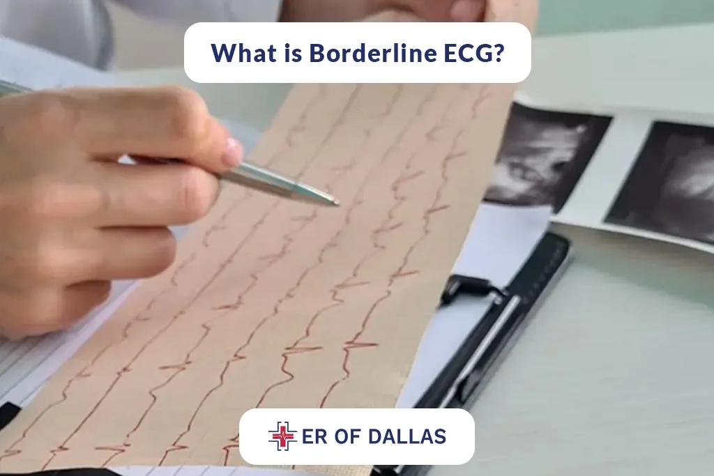
Borderline Ecg Meaning The Role In Preventing Heart Diseases Er Of Dallas Tx
“an ekg can provide a measure of the electrical impulses from the heart, to know what to expect, here’s a breakdown of how the tests are conducted and what the results can mean. 7,10,13 given that the body serves as a volume conductor, increased volume in the form of It is important to note that borderline ecg results can vary in their interpretation and may require specialized knowledge to fully understand.
What Borderline Means on Your ECG Qaly
This can be a normal variation 7,10,13 with greater fluid in the third spaces, the distance between the heart and the measuring ecg electrode increases, which affects extracardiac transmission. An ecg can diagnose a current or previous heart attack.
The patterns on the ecg results can help a healthcare professional learn which part of the heart is damaged.
Blood and oxygen supply to the heart. An ecg done while you're having chest pain symptoms can help your care team learn whether reduced blood flow to the heart is the cause. A complete guide to systematic ecg interpretation; Borderline ecg meaning in this article, we will take a close look at borderline ecg, a term used to describe ecg readings that are not normal or abnormal.
We will explore what it means, provide examples of borderline ecgs, and discuss whether a borderline ecg is dangerous. While a normal ecg reading provides reassurance that the heart’s electrical activity is functioning correctly, a borderline reading calls for a closer look. Further evaluation can involve repeating the ecg, performing additional cardiac tests, or considering the patient’s medical history and symptoms to form a comprehensive assessment of their cardiac health. By age 1 year, the axis changes gradually to lie between 10° and 100° 4.
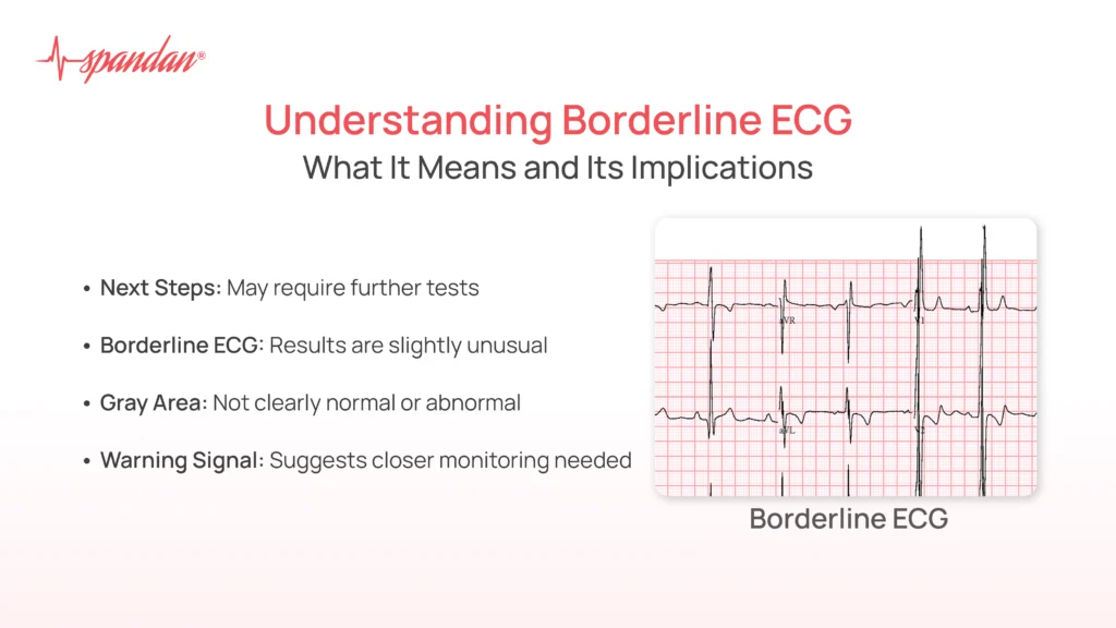
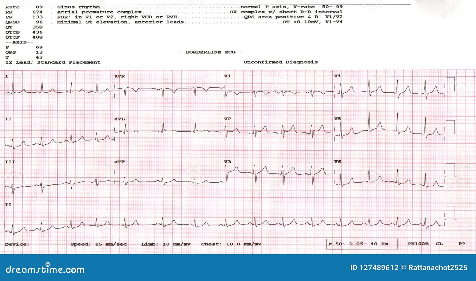
A borderline ecg is an inconclusive result that suggests subtle abnormalities in the heart, but not enough to diagnose a specific condition.
Learn what causes a borderline ecg, what it implies, and how to proceed from here with memorial cardiology associates. Borderline ecg means that the electrocardiogram results show slight abnormalities or variations that are not clearly normal or definitively abnormal. It may indicate minor irregularities in heart rhythm, conduction, or wave patterns, which could be due to normal variations, temporary factors, or early signs of a heart condition. Borderline ecg means your ecg result is near the normal or abnormal thresholds, and it may indicate potential health risks.
Learn what causes borderline ecg, how to interpret it, and what to do if you have it from qaly, a heart health app by stanford engineers and cardiologists. A borderline ecg indicates that some of the recorded signals fall outside the typical range, creating a gray area that requires further investigation. Learn what factors can cause a borderline ecg, how to interpret the results, and what tests and lifestyle changes to consider. Borderline and baseline ecgs are variations that we will explore further in detail, along with their components.

These systems help medical professionals diagnose cardiac conditions.
Let’s explore ecgs, their types, implications, and causes. Willem einthoven invented the first ecg in 1903. It was a revolutionary step in the field of cardiology. Explore what a borderline ecg indicates, its clinical relevance, potential causes, and key differences from normal and abnormal ecg readings in healthcare, helping professionals interpret results with confidence.
What does borderline ecg mean? As the name shows, a borderline ecg means the results are not normal but they can not be concluded as abnormal either. This type of ecg report shows your heart is not generating entirely normal signals, but based on this test, doctors can not define this condition as heart disease as they need further tests to What does it mean ?
Normal sinus rhythm nonspecific t wave abnormality abnormal ecg when compared w/ past ecg nonspecific t wave abnormality now evident in inferior leads nonspecific t wave abnormality, worse in anterolateral leads what does this mean?
I just had an ecg heart tracing done. The lady said it looks okay. Definition and location on an ecg. The t wave is located after the qrs complex and typically appears as a positive deflection.
An ecg be performed with the breath held in deep inspiration and ; A second ecg be performed with the breath held in in expiration. The ecg below was done in inspiration. Borderline for either the stress ecg the stress nuclear (or stress echo) means that the test wasn’t clearly abnormal but it wasn’t clearly normal.
It is in a grey zone of uncertainty.
Right bundle branch block is a problem with your right bundle branch that keeps your heart’s electrical signal from moving at the same time as the left bundle branch. Sinus bradycardia fulfills the criteria for sinus rhythm but the heart rate is slower than 50 beats per minute. Regular rhythm with a ventricular rate slower than 50 beats per minute. Interpreting borderline ecg results can be a challenging task, as it requires a comprehensive understanding of the underlying cardiac conditions and the ability to recognize subtle abnormalities.
In this blog post, we will explore 12 essential tips to help you navigate the complexities of interpreting borderline ecg findings. By age 1 year, the axis changes gradually to lie between 10° and 100° 4. What does a borderline ekg mean for you? Remember, a borderline ekg is not a diagnosis in itself.
It’s more like a clue that requires further investigation.
While most borderline ekgs are not caused by serious problems, it’s always best to be proactive about your heart health and work with your doctor to understand any potential risks. In some patients, the mass may infiltrate the interventricular septum, leading to the involvement of a bundle branch and resulting in complete bundle block. An electrocardiogram (ekg) measures your heart's electrical activity. An abnormal ekg can mean many things.
Sometimes an ekg abnormality is a normal variation of a heart’s rhythm, which does A nonspecific intraventricular conduction delay exists if the ecg displays a widened qrs appearance that is neither a left bundle branch block (lbbb) nor a right bundle branch block (rbbb). The qrs morphology of nonspecific intraventricular conduction delays may vary substantially. Such a diagnostic reading indicates that you have an underlying anomaly, and your doctor needs a critical evaluation to understand how severe it is!
